Incucyte® Base basic software module for cell analysis
Incucyte® Software Workflow
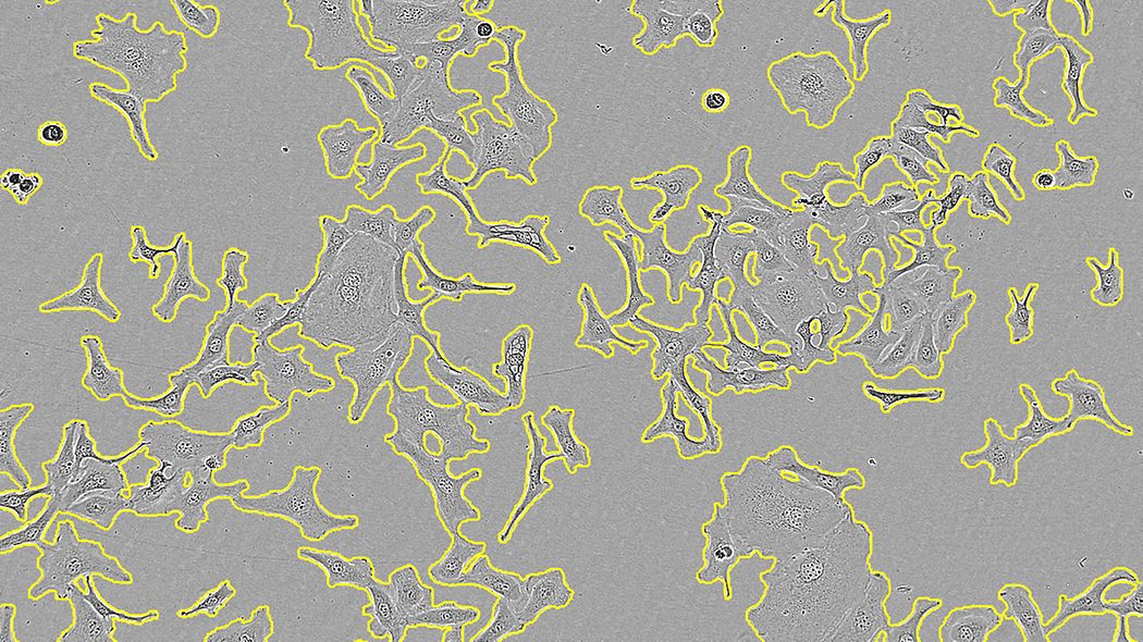
Incucyte® AI Confluence Analysis Workflow

AI-driven confluence analysis provides a simple workflow for highly accurate cell segmentation in phase contrast images, accommodating a wide range of cell types and morphologies with minimal user input. AI confluence analysis is available with Incucyte® v2022A software.
Incucyte® Classic Confluence Analysis Workflow

Classical confluence analysis allows you to tailor phase segmentation parameters to individual cell types and experimental conditions to accurately distinguish background and positive fluorescent objects over a wide range of intensities. Depending on the optical module, it allows the quantification of green, orange, red and/or NIR fluorescent objects within each image.
Overview of Incucyte® software features
Intuitive interface
The Incucyte® Acquisition interface guides users through experimental setups using microplates, flasks and dishes of your choice. Simply select parameters and press "Start" for seamless, continuous live cell imaging.


Simple navigation and image viewing
Incucyte® VesselView enables users to navigate and explore thousands of accelerated images with unprecedented efficiency. By viewing images of all locations in your experiment at once, you can quickly assess treatment effects or identify outliers. You can also overlay metrics to quickly assess and verify image processing parameters. Then, generate presentation-ready images and movies in just a few clicks.

Advanced analysis
The Incucyte® Analysis interface enables even beginners to turn images into powerful insights. Providing dedicated tools to answer your scientific questions, image processing and analysis is uncomplicated yet extremely powerful. Create analysis definitions once, then reapply them to subsequent experiments to generate real-time metrics that enable decision-making. Easily review all trends in an experiment at once with Incucyte® Microplate Graphs and create custom curves that are ready for publication and presentation.


Additional software modules
you might be interested
No posts available